
UIWSOM Student Doctor Creates 3-D Printed Anatomically Accurate Model for Medical Education
By Devin Castillo
T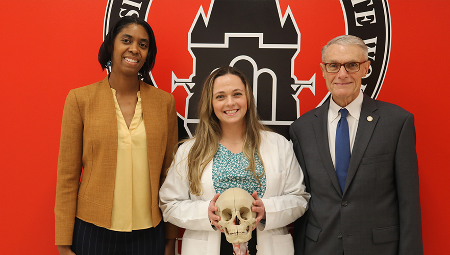 o transform learning communities with the ability to impact civic engagement, education, health and health care in the global community.
This is the vision of the UIW School of Osteopathic Medicine (UIWSOM). With each generation of aspiring healthcare practitioners that enter their doors, UIWSOM commits to improving the health and well-being of our local, national and global communities. “We bring together world-class researchers and faculty from diverse health-related fields to train our future osteopathic physicians,” shared Dean John T. Pham, DO. “Promoting culturally, linguistically and community-responsive care for all patients to enhance patient safety and improve patient outcomes are core principals of our mission.”
Within their brief yet pivotal four years of medical school, each student dedicates themselves to becoming the next pair of capable healing hands that will serve others and continuously consider how to better care for the world with its current and growing needs.
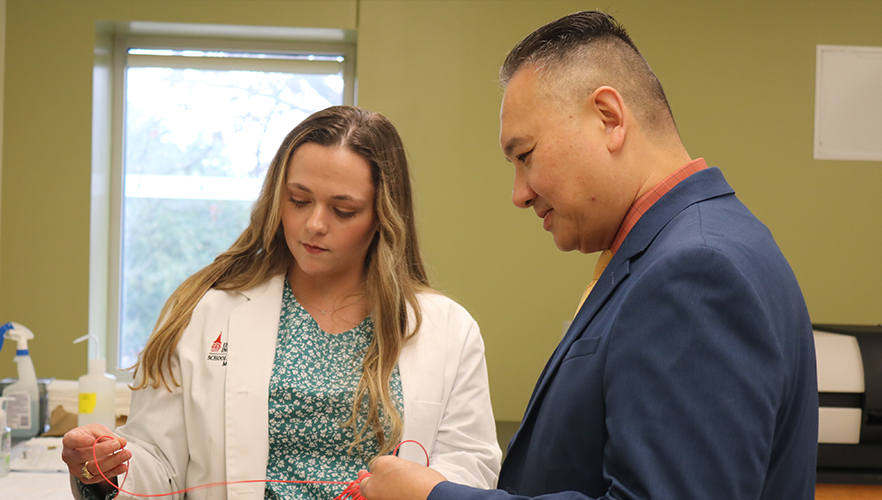
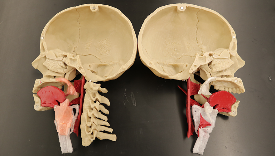
One of these remarkable students is Erin Kraus, a third-year UIWSOM student intending to specialize in Otolaryngology Head & Neck Surgery. While pursuing her master's degree in Cell Systems and Anatomy at the University of Texas Health Science Center at San Antonio (UT Health San Antonio), her thesis centered around building a 3-D model of the human head and neck, specifically the larynx, pharynx and skull, for anatomy education.
Her mentors, Dr. Rekha Kar and Dr. Alan Sakaguchi, tasked her with creating this model to improve anatomy education for health professions students. Although learning 3-D printing required a great deal of work and patience, Kraus was eager to see how her model could come to fruition through a process not previously familiar to her.
“The biggest thing that drove me through the learning process (of 3-D printing) was that this was a step towards implicating a clear way to teach anatomy through a model,” explained Kraus.
“Since 2020, I was inspired by my love for anatomy and was motivated by my ability to create something that could be a teaching aid to not only anatomy and medical students but also patients, residents and providers.”
Her days of learning and practice led to the production of her first model of the human head and neck that aided in the successful defense and acceptance of her master's thesis.
Upon graduating from the program, she was granted permission to further advance the model as a medical student to be used for anatomy education and use in clinical and patient education.
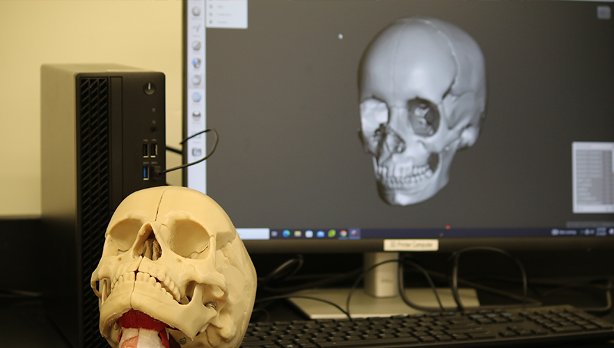
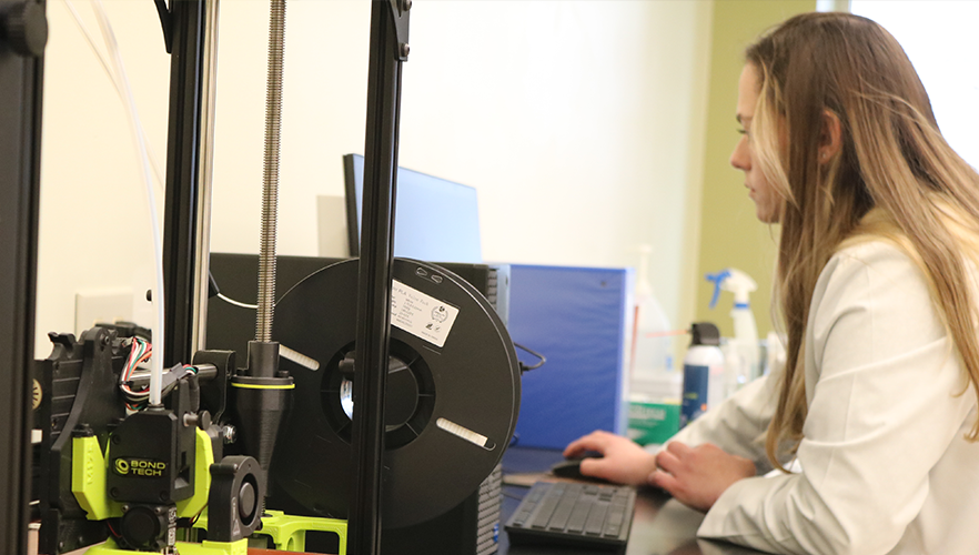
UIWSOM Senior Associate Dean of Academic Affairs Dr. Earlanda Williams served on Kraus’ thesis committee and recruited her to join the UIWSOM family in 2021. Kraus began by developing a virtual model using computer-aided design (CAD) software. She then imported the model into the 3-D printer’s slicing software where it was sliced into thousands of layers. Finally, she oversaw the printer utilizing fused deposition modeling (FDM) to build the model from the foundation up using layers upon layers of melted plastic.
Kraus first started off at UIWSOM as an employee, where she was a part of the structures team serving as an anatomy teaching laboratory assistant. For a year, she was responsible for dissecting and preparing cadavers for UIWSOM's human gross anatomy courses. In 2022, she officially began her medical studies as a UIWSOM student.
“At UT Health, I collaborated with the school Librarian who completed the printing of my model. At UIWSOM, I had the opportunity to personally create the model from start to finish. It was a bit daunting as I trialed and errored everything on my own.”
As she better familiarized herself with 3-D printing independently, she considered how to enhance the aesthetics and functionality of her head and neck model. She tailored it to have specific features that allowed for use in clinical training. For example, she added silicone for the mucosa of the upper airway and larynx, which could be used for procedural and clinical training.
Kraus hopes that her model will aid not only medical students in understanding the anatomy of the head and neck but also clinicians, residents and patients. These models have the potential to offer reliable visual aids to help patients’ comprehension of their clinical procedures and provide artificial subjects compatible with medical demonstrations.
Additionally, she hopes her model will offer an anatomically accurate educational resource as an alternative to cadaver-based anatomy education, and for underserved schools without access to traditional gross anatomy educational resources. Kraus intends for the 3D files for her model to be offered as “open source,” meaning other institutions could freely access her coding to create copies of her head and neck model for themselves.
Although the model will require access to a 3-D printer to be created, it only takes an average low cost of $30 in materials to build. This will allow other medical institutions to acquire a cost-effective resource that doesn’t require the costs and laboratory equipment needed for a human gross anatomy lab, as the models are made from completely artificial materials.
“I think there were many extra things that I hadn’t originally anticipated with the model, but it was really rewarding,” admitted Kraus. “To see the model come to life in front of my eyes with all its advances were fascinating. Understanding your patient's anatomy is important when treating them. Having this tool would be immensely helpful because we could tailor the model to specific teaching goals.”
In 2024, Erin was also given the opportunity to return to UT Health San Antonio to present on the recent state of her model to the Department of Otolaryngology Head and Neck Surgery, highlighting how she has given significant consideration to the development of the model so it can be an effective tool across various platforms, and sharing her hopes for how the model will be utilized in the future. It was a moment to demonstrate to her alma mater the progress made on her research and make her present UIW mentors proud.
"It has been an immense pleasure to mentor and support student doctor Erin Kraus in her anatomy research endeavors since her time as a graduate student at UT Health,” expressed Williams. “Ms. Kraus has displayed resounding innovation, dedication and resilience as she initiated this project during the early months of the COVID-19 pandemic and established opportunities to collaborate with UIWSOM to complete her project. Erin has continued to thrive as an anatomy educator and osteopathic medical student over the past four years. I am both proud and confident that student doctor Erin Kraus will continue to make significant contributions to the field of anatomy and medical education for many years to come."
"Student doctor Erin Kraus is an exemplary role model for professionalism, integrity and dedication to excellence that characterizes the physician of the future, and the mission of the UIW School of Osteopathic Medicine,” remarked Dr. Richard Holt, professor of Clinical and Applied Science Education who introduced Kraus to the field of Otolaryngology. “Her knowledge of human anatomy and the requisite ingenuity for developing open access, 3-D printed, anatomically correct models that will be available globally for medical education, particularly in developing countries, present an outstanding and altruistic educational advancement. Ms. Kraus' goal to become an otolaryngologist head and neck surgeon, is certainly one I fully support. I project that Ms. Kraus will continue to contribute to medical education, medical knowledge, simulation surgery advances and clinical innovations in her future academic career."
Kraus never imagined that this model would follow her throughout her educational career, but she is grateful for the opportunities it has offered and the people that it has unexpectedly ushered into her life. She is deeply grateful to UIWSOM and her mentors Dr. Earlanda Williams, Dr. Richard Holt, Dr. Yolanda Rangel and many more. She recognizes how everyone has believed in her, offering her an abundance of support, encouragement and guidance throughout these past few years.
Because of their support, Kraus has been able to develop several other novel projects involving 3-D printed models to support her peers and help them learn how to apply basic anatomy to complex clinical cases. An example of one of these projects is the printing of miniature, pocket-sized pelvis models that can be manipulated and used to demonstrate the biomechanics and positions of the pelvis.
These were designed to help osteopathic medical students with understanding and demonstrating somatic dysfunction of the pelvis. “My number one priority is to be a doctor,” assured Kraus. “I hope that this head and neck model can follow me throughout my career and that I can expand further on it as I continue to become a physician. If I can incorporate the model in my training right now, that would be great. At the end of the day, though, I want to learn how to be a good doctor and help others to become good doctors.”
It is through the innovation, compassion and unwavering dedication of students that UIWSOM can see their vision come to life. Although students only spend a brief time at the school, it is undeniable that as they move beyond the Nest, they will be active contributors to a better world for all.